


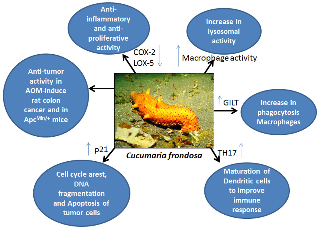
Inflammation reflects a sequence of events that occurs in response to injury or foreign body entry; this process is highly coordinated and involves various cell types. A normal inflammatory response is characterized by the infiltration of leukocytes and release of other activated inflammatory mediators at the injury/infection site, and eventually will be resolved or regulated with the release of anti-inflammatory mediators. This action is needed to restrict the ongoing inflammation and stop its development into chronic inflammation. Persistent, long-lasting inflammation may lead to inflammatory disease and, eventually, cancer. Although several anti-inflammatory agents, like non-steroidal anti-inflammatory drugs (NSAIDs), are available, their use is restricted in dosage or intervals and special precautions are advised due to their gastrointestinal toxicity. Therefore, there is a need for the development of natural anti-inflammatory agents that have the potential to self-limit or resolve inflammatory events, without progressing into chronic inflammation.
Sea cucumber extracts are reported to affect soft tissue repair. Different sea cucumber extracts display different functions, with varied effects when tested in vitro or in vivo. Compared with the high dose (10 mg/kg), low-dose Stichopus sp1 extract (1 mg/kg) was shown to promote healing properties in rabbits with fractures; these findings indicate that doses are important, and these compounds may not produce dose-dependent effects. The major fatty acids in sea cucumber extracts, EPA and DHA, contribute to tissue/wound healing. EPA and DHA are known n3 fatty acids, which inhibit prostaglandin synthesis by suppressing COX-2 and 5-LOX expression under inflammatory conditions, and also act as anti-thrombotics, which accelerate the healing process. These tissue repair properties might play a role during tumor development, when the unique fatty acid content may help to inhibit cancerous transformation of epithelial cells.
Patents by Collins provide wide information on the possible use of bioactive components of sea cucumber tissue fractions as preventive or therapeutic agents to curb inflammation. The anti-inflammatory properties of Australian sea cucumbers with a sea plant formulation (95% w/w H. (Microthele) nobilis, H. (Microthele) axiologa and Stichopus variegatus) and sea plant (5% w/w Sargassum pallidum) have been well demonstrated in male Dark Agouti or hooded Wistar rats. Although this formulation produced hypotensive side effects, a dose of 50 mg/kg (ip) was equally effective in inhibiting paw inflammation when compared with 150 mg/kg of aspirin. Holothuria tubulosa, Leptogorgia ceratophyta, Coscinasterias tenuispina, and Phallusia fumigata extracts were shown to be effective in down regulating pro-inflammatory marker cyclooxygenase (COX) activity in inflamed mouse tissues. As it is well documented that COX-2 is involved in carcinogenesis, sea cucumber extracts should be further analyzed for their anti-cancer properties.
Heparin analogues obtained from the sea cucumber Styela plicata produced anti-inflammatory effects in a rat model of colon inflammation. Subcutaneous administration of GAGs (4 or 8 mg/kg per day) over seven days reduced colitis induced by 2,4,6-trinitrobenzene sulfonic acid (TNBS) in a Wistar rat. Furthermore, treatment group animals showed less infiltration of macrophages and T-cells, and profoundly reduced the TNF-α and other signaling pathways, including the NF-κB and MAP kinase pathways. No hemorrhagic events were observed with the administration of GAGs. This report suggests that GAGs produce anti-inflammatory and immune response effects in a rat model of colitis. Further analysis of these compounds for their utility in colon cancer prevention and treatment is warranted.
The dermatan sulfates from S. plicata (sulfated at carbon 4) and P. nigra (sulfated at carbon 6), which contain the same disaccharide core structure [α-l-IdoA(2SO4)-1→3β-d-GalNAc]n, are reported to be potent inhibitors of P-selectin. In a thioglycollate-induced mouse model of peritonitis, dermatan sulfates showed drastic inhibition of metastasis of MC-38 colon carcinoma and B16-BL6 melanoma cells, regardless of the position of sulfation on the N-acetylgalactosamine, and also decreased the infiltration of inflammatory cells. Fucosylated chondroitin sulfate (fCS) isolated from the sea cucumber H. forskali showed high affinity binding of the oversulfated CS/DS chains containing GlcAβ(1→3)GalNAcβ4,6S disaccharide units to L- and P-selectins. In addition, fCS isolated from this sea cucumber inhibited neutrophil migration.
Post time: Jan-01-2024